Institute of Cell Biology · University of Bern

Cell Fate & Nuclear Organization

How is an entire genome folded inside a micron-scale nucleus, and how does three-dimensional chromosome architecture shape transcription, cell identity, animal behaviour and organismal aging? Using Caenorhabditis elegans, we combine classical genetics, genome engineering, molecular biology and live imaging to dissect genome organization in vivo.

6 SMC Motors Specialized somatic condensins & cohesins folding holocentric chromosomes
~38 kb Fountains CohesinCOH-1 3D enhancer loops regulating neuronal isoforms & behavior
2-Cell Resolution Tissue-specific RAPID & DamID genomics in intact living nematodes
C. elegans genome-wide Hi-C contact map with fluorescent nematodes
Holocentric 3D Genome Architecture in C. elegans Whole-genome Hi-C contact matrix (chrI–chrX) & live nematode fluorescence
Inspect ↗
Chromosome X and V DNA-FISH territories in C. elegans embryos
Chromosome X & V Territories DNA-FISH
Subnuclear SDC bodies imaged by spinning-disk microscopy
Nuclear SDC Bodies Nat Genet '24

Folding the Genome Across Scales & Cell Types

Nearly every cell in an animal carries the exact same DNA blueprint, yet executes a distinct transcriptional program inside a 5-micrometer nucleus. We ask how molecular motors, nuclear landmarks, and chromatin states organize this 3D space—and what happens to development, physiology, and behavior when that architecture is rewired.

Packing two meters of human DNA inside a microscopic nucleus is equivalent to folding a fine wire stretching from Zürich to Geneva inside a basketball. Far from being homogeneous, this folding segregates active and silent regions between the nuclear lamina, nuclear pores, and subnuclear condensates, while Structural Maintenance of Chromosomes (SMC) motor complexes actively extrude DNA loops across kilobase to megabase scales.

We use the roundworm Caenorhabditis elegans—with its invariant cell lineage, transparent body, 3-day life cycle, and holocentric chromosomes lacking the vertebrate insulator CTCF—as an in vivo system where 3D chromatin topology can be perturbed post-mitotically and read out directly at the level of single identified neurons, tissues, lifespans, and animal behaviors.

Our recent work revealed a striking evolutionary division of labor: whereas mammals rely on cohesin for megabase TADs, C. elegans uses condensin I for long-range autosomal folding, a specialized condensin IDC and nuclear SDC bodies for X-chromosome dosage compensation, and interphase cohesinCOH-1 to extrude ~38 kb enhancer fountains essential for neuronal gene regulation.

Multi-scale C. elegans interphase genome organization
Interphase nuclear architecture from whole-chromosome territories (T/C domains) to enhancer fountains and SDC bodies · Erkan, Ghadage & Meister, Curr Opin Genet Dev (2026) Zoom ↗

Current Research Axes

Click any figure to inspect high-resolution data
3D Genome Folding Hi-C contact maps of C. elegans Chromosome II and Chromosome X Hi-C Maps ↗

Holocentric Chromosome Folding by Condensin I

By engineering TEV protease-cleavable kleisins across all five somatic cohesin and condensin complexes in post-mitotic larvae, we demonstrated that condensin I—rather than cohesin—is the primary long-range (100 kb–3 Mb) loop extruder in C. elegans. Cleaving condensin I triggers genome-wide decompaction, chromosome mixing, and reinforcement of megabase T/C domains and fine-scale (2–40 kb) A/B chromatin compartments.

Enhancer Fountains & Behavior SKN-1::GFP isoform switching in ASI head sensory neurons upon cohesin cleavage Locus + ASI Neurons ↗

Enhancer “Fountains”, Neuronal Isoform Choice & Cohesinopathies

How do 30,000+ nematode enhancers achieve target specificity without autosomal TADs? Using 2-kb Hi-C and bTMP supercoiling profiling, we uncovered 935 ~38 kb chromatin fountains extruded by interphase cohesinCOH-1 at active, topoisomerase-bound enhancers. Fountain collapse derepresses neuronal genes, switches skn-1/Nrf promoter usage in ASI sensory neurons, and impairs foraging behavior—mirroring human Cornelia de Lange cohesinopathies.

Dosage Compensation Spinning-disk microscopy of subnuclear SDC bodies on the X chromosome SDC Bodies ↗

X-Chromosome Topology, Nuclear Pores & Subnuclear SDC Bodies

To equalize X-linked gene dosage between XO males and XX hermaphrodites, the dosage compensation complex (DCC) recruits condensin IDC via direct binding of SDC-3 to the DPY-27 elbow coiled-coil, unlocking DNA loop extrusion to fold X-specific TADs. Multi-way Hi-C and live imaging revealed that 35 rex TAD boundaries self-assemble into subnuclear SDC bodies that safeguard repression and lifespan.

Nuclear Envelope, Cell Fate & Genomics Two-color fluorescence microscopy of neuroendocrine XXX cells discovered by RAPID RAPID Microscopy ↗

Nuclear Envelope Kinases, Chromatin Remodeling & Single-Tissue Footprinting

We dissect how nuclear periphery regulators and chromatin remodelers govern cell fate, stress recovery, and disease—including H3K9me3 ChromID proteomics and VRK-1–BAF-1 chromatin–lamina release, decoupling mitotic vs. motor-neuropathy roles of VRK-1, poised LET-418/Mi-2 immune control, Polycomb/Notch plasticity, and 2-cell RAPID footprinting.

Live Imaging & Experimental Snapshots

From four-dimensional DIC embryogenesis recordings and two-color chromosome painting to high-resolution Hi-C matrices and quantitative behavioral tracking across our publications.

Live DIC Time-Lapse · Embryogenesis

Synchronous Early Development of C. elegans Embryos

C. elegans develops from a fertilized zygote into a 558-cell freshly hatched larva through a completely invariant, rapid cell lineage. As blastomeres divide and commit to muscle, epidermal, neuronal, and intestinal fates, chromosomes transition from plastic embryonic organization into cell-type-specific 3D nuclear territories and dosage-compensated X domains.

Invariant Lineage 30–40-Cell DCC Onset Live Whole-Animal Readouts
VRK-1::wrmScarlet and HIS-72::GFP mitotic nuclear rim and anaphase bridges in C. elegans embryos

VRK-1 Mitotic Bridges & Neuropathy Motor Defects

Spinning-disk imaging captures VRK-1::wrmScarlet nuclear rim remodeling and anaphase chromosome strands, while BAB-patch mutations decouple progressive motor dysfunction from mitosis.

Spinning-Disk · VRK-1 microPubl Biol 2026 →
2-color confocal DNA-FISH chromosome territories in C. elegans embryos

Whole-Chromosome Painting & Single-Locus DNA-FISH

Isothermal Phi29 rolling-circle amplification generates high-density ATTO-488 and ATTO-647N probes resolving single 12 kb integrations and discrete Chr X / Chr V territories in embryos.

Confocal · DNA-FISH STAR Protocols 2020 →
Stage-specific chromatin fountains at active enhancers

Stage-Specific Chromatin Fountains at Active Enhancers

2-kb resolution Hi-C pileups reveal symmetric and asymmetric ~38 kb loops extruded bidirectionally from active embryonic and larval enhancers by cohesinCOH-1.

Hi-C · bTMP-seq Nat Commun 2026 →
X-chromosome rosette loop anchors upon condensin cleavage

X-Chromosome Rosette Hubs & Multi-Way Contacts

Removing condensin I/IDC uncovers long-range reciprocal contacts (>8 Mb) and three-way interactions across 35 strong rex sites colocalizing with subnuclear SDC bodies.

MC-Hi-C · X Rosette Nat Genet 2024 →
C. elegans foraging trajectories after cohesin cleavage

From 3D Enhancer Topology to Foraging Behavior

High-content Tierpsy behavioral phenotyping across 47 postural and motion features links interphase cohesinCOH-1 fountain integrity in neurons to food-search navigation.

Tierpsy · Neurobehavior Nat Commun 2026 →
Single-cell transdifferentiation plasticity sensor in C. elegans

Single-Cell Fate Challenge in Intact Living Larvae

Inducible MyoD/GATA-1 challenge in L1 larvae highlights extraordinary cell-fate lock-in across somatic lineages, with selective competence in the saddle-shaped anal sphincter cell.

Spinning-Disk · Plasticity Life Sci Alliance 2019 →
Intestinal pals-5p::GFP innate immune activation in let-418 mutants

Intestinal DamID & Pathogen Defense Chromatin

Cre/lox intestinal DamID-seq reveals LET-418/Mi-2 and MEP-1 poise innate immune pals genes in a repressed yet inducible chromatin state activated via ZIP-1 upon infection.

DamID-seq · Immunity BMC Genomics 2025 →

Publications & Preprints

Current preprints and highlighted recent discoveries from the Meister Lab, followed by our searchable archive with direct PDF downloads, DOIs, and PubMed records.

Current Preprints bioRxiv · 2025–2026

Click schematic to inspect full summary figure ↗
HPL-2 chromoshadow dimerization bridges and segregates H3K9me3 chromatin
bioRxiv · Nov 2026 (v2)Preprint

HP1 dimerization drives the condensation and segregation of H3K9me-marked chromatin

Eswara K., Semple J.*, Rivas-Cuestas F.*, Muzzopappa F., et al., Erdel F., Meister P., and Fischle W. Establishes HPL-2 dimerization as the core driver of H3K9me chromatin condensation and uncouples HP1 foci from gene silencing in vivo.

Mitochondrial stress signaling via PMK-3 and CBP-3 reshapes nuclear heterochromatin upon MRG-1 loss
bioRxiv · August 2026Preprint

Mitochondrial stress signaling shapes the nuclear response to loss of the chromatin reader MRG-1

Zaratiegui C., Rezende Pabst F., Rodriguez Palero M.-J., Meister P., Artal-Sanz M., and Cabianca D.S. Reveals PMK-3/MAPK–CBP-3 mitochondrial stress signaling as an adaptive driver of heterochromatin detachment and gene regulation upon mrg-1 loss.

H3K9me3 ChromID and VRK-1–BAF-1 perinuclear chromatin regulation
bioRxiv · July 2026Preprint

Proximity labeling at H3K9me3 reveals VRK-1 regulate global chromatin distribution in C. elegans

Smith W., Aksianiuk V., Pfaendler R., Villaseñor R., et al., Meister P. Maps the H3K9me3 proteome via ChromID and uncovers VRK-1 phosphorylation of BAF-1 as essential for releasing perinuclear chromatin after stress.

Cryo-EM recruitment of condensin IDC via SDC-3 binding to DPY-27 elbow
bioRxiv · March 2026Preprint

Structural basis of condensin recruitment for X chromosome repression

Valdes Gutierrez A.*, Amudhan G.*, Bernasconi D., Erkan S., Hassler M., Suter I., et al., Meister P., and Haering C.H. Cryo-EM resolves SDC-3 bound to the DPY-27 elbow of condensin IDC, linking X recruitment and loop extrusion to dosage compensation.

Highlighted Publications

Selected peer-reviewed articles (2020–2026)
2026Preprint
HP1 dimerization drives the condensation and segregation of H3K9me-marked chromatin
Eswara K., Semple J.*, Rivas-Cuestas F.*, Muzzopappa F., Ali S., El Mouridi S., Ostwal Y., Frøkjær-Jensen C., Erdel F., Meister P., and Fischle W.
bioRxiv preprint (v2), 2025.11.13.687974
2026Preprint
Mitochondrial stress signaling shapes the nuclear response to loss of the chromatin reader MRG-1
Zaratiegui C., Rezende Pabst F., Rodriguez Palero M.-J., Meister P., Artal-Sanz M., and Cabianca D.S.
bioRxiv preprint, 2026.08.14.744963
2026Preprint
Proximity labeling at H3K9me3 reveals VRK-1 regulate global chromatin distribution in C. elegans
Smith W., Aksianiuk V., Pfaendler R., Villaseñor R., Siva Sankar D., Stumpe M., Lenart P., Askjaer P., Towbin B.D., Baubec T., Dengjel J., and Meister P.
bioRxiv preprint, 2026.07.07.737140
2026Preprint
Structural Basis of Condensin Recruitment for X Chromosome Repression
Valdes Gutierrez A.*, Amudhan G.*, Bernasconi D., Erkan S., Hassler M., Suter I., Wilde B., Bender J., Warscheid B., Meister P., and Haering C.H.
bioRxiv preprint, 2026.03.13.711519
2026
Human disease-relevant mutations in the VRK-1 basic-acidic-basic patch result in motor impairment independent of mitotic defects in C. elegans
Smith W. and Meister P.
microPublication Biology, 10.17912/micropub.biology.002279
2026
Chromosome organization by Structural Maintenance of Chromosomes complexes in C. elegans
Erkan S.*, Ghadage K.*, and Meister P.
Current Opinion in Genetics & Development 96, 102430
2026
Cohesin forms fountains at active enhancers in C. elegans
Lüthi B.N., Semple J.I., Haemmerli A., Thapliyal S., Ghadage K., Stojanovski K., D’Asaro D., Das M., Gilbert N., Glauser D.A., Towbin B., Jost D., and Meister P.
Nature Communications 17, 681
2025
The chromatin remodeler LET-418/Mi-2 regulates the intracellular pathogen response in the C. elegans intestine
Rajopadhye S., Lažetić V., Rodriguez-Crespo D., Troemel E., Meister P., and Wicky C.
BMC Genomics 26, 938
2024
Condensin I folds the Caenorhabditis elegans genome
Das M., Semple J.I., Haemmerli A., Volodkina V., Scotton J., Gitchev T., Annan A., Campos J., Statzer C., Dakhovnik A., Ewald C.Y., Mozziconacci J., and Meister P.
Nature Genetics 56, 1737–1749
2022
3DPolyS-LE: an accessible simulation framework to model the interplay between chromatin and loop extrusion
Gitchev T., Zala G., Meister P., and Jost D.
Bioinformatics 38(24), 5454–5456
2021
Tissue-specific DamID protocol using nanopore sequencing
Gómez-Saldivar G., Glauser D.A., and Meister P.
Journal of Biological Methods 8(3), e152
2020
Tissue-specific transcription footprinting using RNA Pol DamID (RAPID) in Caenorhabditis elegans
Gómez-Saldivar G.*, Osuna-Luque J.*, Semple J.I., Glauser D.A., Jarriault S., and Meister P.
Genetics 216(4), 931–945
2020
Generation of Inexpensive, Highly Labeled Probes for Fluorescence In Situ Hybridization (FISH)
Sharma R. and Meister P.
STAR Protocols 1(1), 100006
2019
Males as somatic investment in a parthenogenetic nematode
Grosmaire M., Launay C., Siegwald M., Brugière T., Estrada-Virrueta L., Berger D., Burny C., Modolo L., Blaxter M., Meister P., Félix M.-A., Gouyon P.-H., and Delattre M.
Science 363, 1210–1213
2019
Polycomb and Notch signaling regulate cell proliferation potential during Caenorhabditis elegans life cycle
Coraggio F.*, Püschel R.*, Marti A., and Meister P.
Life Science Alliance 2(1), e201800170
2017
Coupling 1D modifications and 3D nuclear organization: data, models and function
Jost D., Vaillant C., and Meister P.
Current Opinion in Cell Biology 44, 20–27
2016
Tools for DNA adenine methyltransferase identification analysis of nuclear organization during C. elegans development
Sharma R., Ritler D., and Meister P.
Genesis 54(3), 151–159
2016
From single genes to entire genomes: the search for a function of nuclear organization
Püschel R., Coraggio F., and Meister P.
Development 143, 910–923
2016
Dosage compensation and nuclear organization: cluster to control chromosome-wide gene expression
Sharma R. and Meister P.
Current Opinion in Genetics & Development 37, 9–16
2015
Linking dosage compensation and X chromosome nuclear organization in C. elegans
Sharma R. and Meister P.
Nucleus 6(4), 266–272
2015
DamID of nuclear envelope proteins in C. elegans
Gómez-Saldivar G., Meister P., and Askjaer P.
Methods in Molecular Biology 1411, 355–369
2014
Differential spatial and structural organisation of the X chromosome underlies dosage compensation in C. elegans
Sharma R., Jost D., Kind J., Gómez-Saldivar G., van Steensel B., Askjaer P., Vaillant C., and Meister P.
Genes & Development 28, 2591–2596
2014
A conserved role for p48 homologs in protecting dopaminergic neurons from oxidative stress
Bou Dib P., Gnägi B., Daly F., Sabado V., Glauser D., Meister P., and Nagoshi E.
PLoS Genetics 10, e1004718
2014
Modern tools to study nuclear pore complexes and nucleocytoplasmic transport in C. elegans
Askjaer P., Galy V., and Meister P.
Methods in Cell Biology 122, 277–310
2014
Modern methods in C. elegans chromatin biology
Askjaer P., Ercan S., and Meister P.
WormBook, 1–35
2013
Promoter- and RNA polymerase II-dependent hsp-16 gene association with nuclear pores in C. elegans
Rohner S., Kalck V., Wang F., Ikegami K., Lieb J.D., Gasser S.M., and Meister P.
Journal of Cell Biology 200(5), 589–604
2013
Nuclear organization in the nematode C. elegans
Sharma R. and Meister P.
Current Opinion in Cell Biology 25, 395–402
2013
Building silent compartments at the nuclear periphery: a recurrent theme
Meister P. and Taddei A.
Current Opinion in Genetics & Development 23, 96–103
2013
Microscopic analysis of chromatin localization and dynamics in C. elegans
Lanctôt C. and Meister P.
Methods in Molecular Biology 1042, 153–172
2012
Step-wise methylation of histone H3K9 positions heterochromatin at the nuclear periphery
Towbin B.D., González-Aguilera C., Sack R., Gaidatzis D., Kalck V., Meister P., Askjaer P., and Gasser S.M.
Cell 150, 934–947
2011
C. elegans HP1 expression profiling links developmental plasticity, longevity, and lipid metabolism
Meister P., Schott S., Rohner S., Gasser S.M., and Palladino F.
Genome Biology 12, R123
2011
Expression of EDMD mutant lamin in C. elegans causes motility defects, muscle disorganization and prevents active gene relocation in muscle cells
Mattout A., Pike B.L., Towbin B.D., Bank E., Meister P., Gruenbaum Y., and Gasser S.M.
Current Biology 21, 1603–1614
2011
Nuclear geometry and rapid mitosis ensure asymmetric episome segregation and rejuvenation in yeast
Gehlen L.R., Nagai S., Shimada K., Taddei A., Meister P., and Gasser S.M.
Current Biology 21, 25–33
2011
Locking the genome: 3D nuclear organization and cell fate acquisition
Meister P., Mango S.E., and Gasser S.M.
Current Opinion in Genetics & Development 21, 167–174
2010
The spatial dynamics of developmentally regulated genes during C. elegans development
Meister P., Towbin B.D., Pike B.L., Ponti A., and Gasser S.M.
Genes & Development 24, 766–782
2010
Repetitive transgenes in C. elegans accumulate heterochromatic marks and are sequestered at the nuclear envelope in a copy-number- and lamin-dependent manner
Towbin B.D., Meister P., Pike B.L., and Gasser S.M.
Cold Spring Harbor Symposia on Quantitative Biology LXXV, 555–565
2010
Visualizing yeast chromosomes and nuclear architecture
Meister P., Gehlen L.R., Varela E., Kalck V., and Gasser S.M.
Methods in Enzymology 470, 535–567
2009
The nuclear envelope — a scaffold for silencing?
Towbin B.D., Meister P., and Gasser S.M.
Current Opinion in Genetics & Development 19, 180–186
2008
Modules for cloning-free chromatin tagging in Saccharomyces cerevisiae
Rohner S., Gasser S.M., and Meister P.
Yeast 25, 235–239
2007
Replication foci dynamics: replication patterns are modulated by S-phase checkpoint kinases in fission yeast
Meister P., Taddei A., Ponti A., Baldacci G., and Gasser S.M.
EMBO Journal 26, 1315–1326
2006
In and out of the replication factory
Meister P., Taddei A., and Gasser S.M.
Cell 125, 1233–1235
2005
Temporal separation of replication and recombination requires the intra-S checkpoint
Meister P., Taddei A., Vernis L., Poidevin M., Gasser S.M., and Baldacci G.
Journal of Cell Biology 168, 537–544
2003
Nuclear factories for signalling and repairing DNA double strand breaks in living fission yeast
Meister P., Poidevin M., Francesconi S., Tratner I., Zarzov P., and Baldacci G.
Nucleic Acids Research 31, 5064–5073

The Meister Lab Team

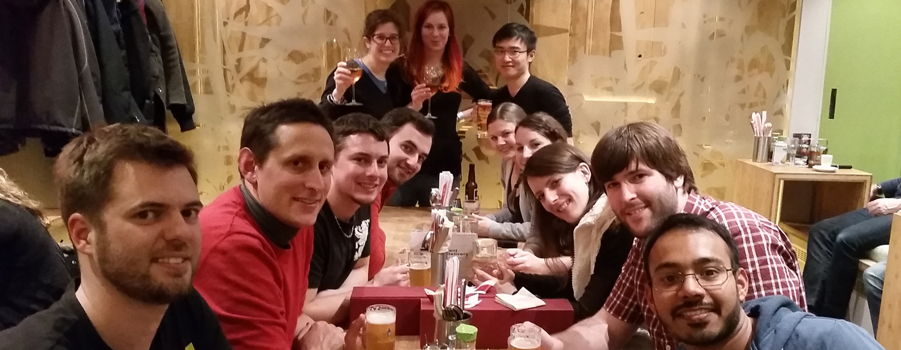
An international team of cell biologists, geneticists, microscopists, and computational biologists at the Institute of Cell Biology, University of Bern.

Meister Lab group photo in Bern, Spring 2026
Cell Fate & Nuclear Organization Laboratory · Spring 2026 Click photo to enlarge ↗

Current Members

Peter MeisterFR

Peter Meister

Group Leader · Prof. Dr.

Jennifer SempleCH / UK

Jennifer Semple

Laboratory Manager & Bioinformatics

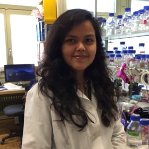
Moushumi DasIN

Moushumi Das

Scientific Assistant

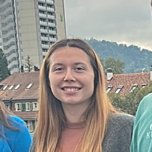
Valeryia AksianiukUS / BY

Valeryia Aksianiuk

PhD Student

Kalyan GhadageIN

Kalyan Ghadage

PhD Student

Dario BernasconiCH

Dario Bernasconi

PhD Student

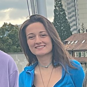
Sinem ErkanTR

Sinem Erkan

PhD Student

Melika Azaripour EsfahaniIR

Melika Azaripour Esfahani

PhD Student

Supported by SBFI / ESKAS

Alumni

NameOriginRole in LabSubsequent Position
Will Smith

UKPhD student
Aleksa Yan
RUVisiting PhD student (SBFI/ESKAS)PhD student, Novosibirsk
Lise Durif
FRVisiting studentStudent, Institut de Sciences Politiques, Paris
ISIris Suter
CHMaster student
AHAnja Haemmerli
CHMaster studentTrainee, IFIK, University of Bern
Jaime Osuna-Luque
ESPhD studentInside Sales Representative, Takara Bio Europe, France
Bolaji Isiaka (-Lüthi)
NGPhD studentProject Leader QC, Lonza, Visp
Valeriia (Lera) Volodkina
RUPhD student
Todor Gitchev
BGMaster studentSenior Research Software Engineer, Krauthammer Lab, Zürich
Ahrmad Annan
FRMaster studentPhD student, Ciriello Lab, UNIL Lausanne
Janik Scotton
CHMaster studentPhD student, Simonini Lab, UZH Zürich
JCJulie Campos
FRLaboratory managerTechnician, InvivoGen, Toulouse
SKSonia Karaz
FRLaboratory managerLaboratory Manager, Feige Lab, Lausanne
RPRingo Püschel
DEPhD studentProcess Technology Senior Engineer, CSL Behring, Bern
RSRahul Sharma
INPhD studentTeam Leader, Myllia Biotechnology, Vienna
NRNiels Rinzema
NLMaster studentPost-doc, Stadhouders Lab, Erasmus MC, Rotterdam
AJAbdulaziz Jaber
SAMSc studentScientist II, Roche Diagnostics, Auw
FCFrancesca Coraggio
ITPhD studentAssociate Researcher, Goncalvez Lab, DKFZ Heidelberg
AMAlisha Marti-Grendelmeier
CHMSc student / injection specialistTechnical Owner Lentivirus & CAR-T, Janssen Vaccines, Bern
TGThomas Gfeller
CHMSc studentSr. Team Lead GMP Operations, Janssen Vaccines
AGAdriana Gonzalez-Sandoval
MXPost-docPrincipal Technical Development Scientist, Genentech, USA
DRDominic Ritler
CHMaster studentScientific Associate, Canton Basel-Landschaft
FDFiona Daly
CHMaster studentPrincipal Scientist, CSL Behring R&D, Bern

Open Positions & Training

We welcome curious researchers interested in chromosome biology, epigenetics, live microscopy, and quantitative genomics in C. elegans.

Undergraduate & Master’s Training

We regularly host motivated BSc and MSc students for research internships and thesis projects combining CRISPR genome engineering, spinning-disk fluorescence imaging, Nanopore DamID/RAPID, and computational genomics. Please contact Peter Meister directly with a short statement of interest and CV.

Email peter.meister@unibe.ch

PhD & Postdoctoral Inquiries

Prospective PhD candidates (affiliated with the Graduate School for Cellular and Biomedical Sciences, GCB) and postdoctoral fellows interested in fellowship applications (EMBO, SNSF, MSCA, ESKAS) are encouraged to reach out early with research ideas, CV, and contact details of referees.

Inquire by Email →
Research Supported By
  • Swiss National Science Foundation (SNSF)
  • University of Bern
  • Novartis Foundation for Medical-Biological Research
  • Uniscientia Foundation
  • SBFI / ESKAS Excellence Scholarships

Get in Touch

Cell Fate & Nuclear Organization Laboratory
Institute of Cell Biology (IZB), University of Bern
Baltzerstrasse 4, CH-3012 Bern, Switzerland
+41 31 631 46 09